| |
the CASE HISTORY ON SURGERY
the Diagnosis: a blastoma of a mammary gland
BLASTOMA GLANDULAE MAMMARIAE
THE REVIEW OF THE LITERATURE
(Tumor, neoplasma, blastoma), aboriginal superfluous, independent, atypical pathological growths of one or several tissues of the organism arising without the seen causes owing to reproduction of cellular elements, the biological properties of cells caused by changes under influence of blastomogenic factors external and an internal environment refer to as tumours, or neoplasms. The doctrine about tumours, or neoplasms, is secured in an independent department of a science under the name an oncology.
In a basis of tumoral growth boundless reproduction of cells which is not coordinated with growth of other tissues of an organism lays and proceeds after elimination of the causes (carcinogenic substances, x-ray and radioactive radiations, traumas, etc.), caused their appearance. Tumor cells are formed of normal cells of any organs and tissues of an organism, As against normal cells they get new special hereditary morphological and functional properties.
Hence, independence and independence of growth (independent growth) both initial atipicheskie morphological and physiological deflections of cellular elements from type of mother cells are the main characteristic attributes of tumours.
Tumours meet at all kinds of animals more often in second half of life, i.e. on the average and old age.
the Etiology and a pathogeny. Various theories of a blastomogenesis are offered many. However the causes of originating of tumours are not found out yet. The most wide-spread up to the last a flying were two theories of their origin: the theory of embrional rudiments Kongejma and the theory of chronic irritation Virhova. These, and also other theories, trying to find the uniform cause for all kinds of tumours, have not received convincing acknowledgement.
Theory Kongejma (dizontogeneticheskaja the theory) reduces an origin of tumours on vegetations superfluous, staying unexpended during the embrional life, switched off of normal communication or the "lost" germinal rudiments. While other cells of the given embrional organism are exposed to division and turn to normal tissues and organs, groups of cells in germinal rudiments remain in a dormant state - a dozing condition. 3ачатки these can be among congenerous him gistologicheski tissues, and also be exposed to moving to tissues alien to them (distopija). When the germ reaches the complete development, embrional rudiments can remain completely latent and anything itself to not show, but they keep in themselves in a potential kind all unexpended high growing capacity which can be shown at any moment under influence of this or that cause (an external irritation or internal infringement of intercellular balance). Then cells are multiplied and effect pullet, embrional, less differentiated (than cells of a normal tissue) cellular elements which sum represents an educing tumour.
Theory Kongejma does not explain, however, all phenomena observable in tumours, originating of tumours in elderly and senile age is especial in malignant neoplasms, and mainly.
According tothe theory of irritation Virhova (irritatsionnaja the theory) the cause of many tumours, especially cancer, is chronic influence on a tissue of physical and chemical irritants - a different kind of traumas, influence of ionizing radiation and a solar energy, chemical substances of an inorganic, organic and biological origin (products of oil refining and coal, some alkaloids, hydrocarbons). The given theory the professional cancer at people and well illustrates animals. For example, a carcinoma cutaneum at bullocks around of horns on a place of affixion of a cord; at horses - a cancer of angles of a mouth as consequence travmatizatsii their iron bit, a carcinoma cutaneum at roentgenologists, a cancer mild at workers on cobaltic mines, a carcinoma cutaneum at workers of a paraffinic industry and working with tar. In a causal relationship with a chronic irritation put also metamorphosis of stomach ulcers, an intestines and a leather into a neoplasm. Practical value of this theory consists that she enables prophylaxis originating of some tumours, however she does not explain the mechanism of transition of normal cells in tumoral, development of a neoplasm when to appearance of her do not precede neither a chronic inflammation, nor influence of external irritants, and also a problem about congenital tumours and about all group of good-quality neoplasms.
the Parasitogenic theory of a blastomogenesisis based on factors of concurrence of originating of tumours with presence in tissues of animals and the person of some parasites: at a large horned stock - the cancer of a liver at a fascioliasis, at dogs and cats - at opistorhoze, at grey rats at a feeding him of oriental cockroaches educes a cancer of tongue and a stomach. The etiological role of parasites in originating a cancer of a liver, apparently, is reduced to a chronic irritation which can promote originating of tumours of a liver on occassion. At the same time it is necessary to note, that at eurysynusic fastsioleznoj invasions in some countries at a large horned stock and spirotserkozah at dogs malignant neoplasms of the conforming organs are not registered in such scales that it was possible to speak about an etiological role of parasites. In many cases experimentally this role of parasites is not proved. Thus, the parasitogenic theory of a cancer is not capable to explain originating of true neoplasms.
The interesting theory of an etiology of tumours isvirus.the Idea that tumours can be caused by agents of a virus nature, for the first time have stated F. Bosc and A.Vogge1 (1903), have experimentally confirmedV.Ellerman and О. Bang (1908), Р. Rous (1911), having established a virus etiology of a leukosis and a sarcoma of hens. Long time counted, that tumoral viruses are the agents exciting porazhaemye by them of a cell to unlimited and noncontrollable reproduction. However L.A.Zilber (1968) has expressed other opinion on tumoral viruses according to which, first, they genetically transform a normal cell in tumoral; second, do not play a role in the further reproduction of already arisen tumor cells and, thirdly, their action essentially differs from a contagious virus.
Supporters of the virus theory consider, that all tumours are invoked by viruses, a role kantserogenov, as well as other irritants, is reduced to building the conditions necessary for realization of action of oncogenous viruses.
Experimental data and clinical observations show, that in some cases crucial importance in originating and development of neoplasms hasa hormonal factor.So, long introduction to mice of oestrogens invoked in them development of a cancer of a mammary gland as at samok, and samtsov. The ovariectomy at a bristle grass vysokorakovyh lines substantially reduces frequency of originating of a tumour of a mammary gland. At castrated twig tumours of a mammary gland are extremely seldom observed, and the castration which has been lead at dogs with a tumour, invokes regression of tumoral growth, a suspension of it, and sometimes complete petering.
the Polyetiological theorydoes not try to reduce all variety of tumours to any one cause of their originating. Any factors - physical, chemical, biological (viruses, parasites) and others, influencing on the genetic apparatus of a cell, invoke a mutation - metamorphosis of normal cellular elements in tumoral.
Thus, from the brief review of the causes of the mechanism of appearance of many forms of tumours it is visible, that they arise from cells of any organs and tissues under influence of those or other factors which nature finally is not found out.
CLASSIFICATION OF TUMOURS
Tumours categorize on morphological characters according to tissues from which they educe. According to it distinguish:epithelial -papillomas, adenomas, cystomas, carcinomas;soedinitelnotkannye -a fibroma, a myxoma, a lipoma, a chondroma, an osteoma, a melanosarcoma;vascular -hemangiomas, lymphangiomas;muscle -myome, rhabdomyomas; froma nervous tissue -gliomy and neuromas;blended -an osteosarcoma, fibromiksohondroma, fibrohondroosteoma. On character of growth and clinical flow of a tumour divide on good-quality and malignant.
Benign tumours. They grow slowly, are surrounded with a capsule more often, do not sprout prilezhashchie a tissue, and move apart them isekspansivnyj growth.In dependence on localization such tumour in one cases can exist during all life of the patient, not causing special harm. In other cases the benign tumour at the growth puts pressure on prilezhashchy an organ, invoking an atrophy, squeezing of vessels and nerves and breaking his function. Benign tumours do not ulcerate, do not give metastasises and do not invoke the general reaction on the part of an organism. The metabolism, structure of blood and the general condition of an animal remain without changes. After removal of a benign tumour there comes complete convalescence (do not recur).
Benign tumours can recur only at their imperfect removal.
Malignant tumours. They will consist of the cells, undergone to irreciprocal pathological changes and the tissues invoking constant progressing intensive growth consisting of these cells. Thus all daughter cells of a tumour appear also changed. Progressing growth of malignant tumours proceeds and after elimination of pristine irritants in connection with an autonomy of their growth.
Prominent feature of cells of malignant tumours - deep morphological deflections from norm: a polymorphism of cells and their nucleus, a multinucleosis, abundance of mitosises, etc. Changes at a ultramicroscopic level Are taped also: reduction of organellas and mitochondrions, set of nucleoluses, the reinforced lamellar complex (apparatus Goldzhi), a vacuolation endoplazmaticheskogo retikuluma, numerous free ribosomes, etc.
Malignant tumours have no capsule and are characterized fast infiltrirujushchim by growth, blasting the next tissues, sprout them. They easily ulcerate. However it is necessary to note, that at animals the phenomena infiltrirujushchego growth of tumours not always show. In some cases malignant tumours (adenokortsinoma a mammary gland at dogs, an angiosarcoma of a spleen) have the expressed borders and it is easy vylushchivajutsja at operative removal. Cells of malignant tumours, growing in lymphatic and blood vessels, can come off both a blood flow and a lymph to be tolerated to other organs and tissues, invoking development of new tumours -metastasises, that is the second feature of malignant tumours.
Development of malignant tumours is accompanied by deep disbolism in an organism of an animal, invoking deterioration of the general condition, a sharp attrition (cachexia) and a dystrophia. This is promoted by specific action of toxins of a tumour, an absorption of the decomposition product, formed as a result of a ulceration and to a consecutive infection, invoke in an animal the general asthenia, a hypochromia anaemia, etc. After operative removal malignant tumours frequently recur with stronger infiltrativnym growth, give regionarnye and the remote metastasises.
Poluzlokachestvennye tumours. They are inclined to infiltrativnomu to growth, but do not form metastasises. Some fibrosarcomas of cats, an alveolar sarcoma and a fibroepithelioma of dogs, some kinds of carcinomas of horses concern to them, etc. an adamantinoma, gemangioperitsitoma,
Division of tumours on good-quality and malignant is conditional and is allowable only at a clinical assessment of disease. So, the benign tumours posed near to the vital organs and breaking their function, at the growth can bring to ruin the patient (a tumour of a brain, a mediastinum, in retroperitoneal space, in polyh or ferruterous organs, etc.).
In some cases benign tumours in the beginning of the growth do not show attributes of malignant neoplasms; and then, being exposed to a malignancy, actually become a transition phase to development of a cancer. Therefore consider, that an animal with a benign tumour alongside with other pathological growths of tissues are in a precancerous condition.
Relapses and metastasises.Malignant tumours are especially dangerous to an organism owing to appearance of relapses and metastasises. Relapses come after removal by surgical way or radial influence when in a tissue have been abandoned or tumoral cellular elements on a place of a former tumour or in the nearest lymph vessels and lymph nodes are not decayed. It meets at malignant tumours with penetrating (infiltrativnym) growth, than at circumscribed sacculated neoplasms more often. Such relapses educe usually or through small time after removal of an initial tumour, or through continue telnye times. It depends on a degree zlokachestvennosti an initial tumour, completeness of keeping of regulations ablastiki and antiblastiki during a surgical oncotomy, a zone of localization of a tumour and its possible communication with regionarnymi lymph nodes, times of a surgical intervention. The earlier the tumour leaves, the it is less than opportunities retsidivirovanija.
Extremely important prominent feature of malignant tumours is their ability to givemetastasises -the secondary (affiliated) tumours educing in various plots of an organism and arising from an initial tumour, but not taking place in immediate communication with it.
In unusual cases metastasises can give and benign tumours. The favorable moments for metastasises - small size of tumor cells and travmatizatsija (even insignificant) tumours.
THE GENERAL INFLUENCE OF TUMOURS ON THE ORGANISM
Benign tumours of any general influence on an organism do not render. They influence him only mestno. Having arisen in a zone of the vital organs and being enlarged, tumours can squeeze tissues of an organ and break function of it. So, benign tumours quite often cause serious complications (a strangulation of an intestines, a bleeding, a thrombosis pologo an organ - an esophagus, intestines, a urethra).
Malignant tumours, it is especial at long existence, render sharply expressed influence on an organism, invoking the general attrition - a cachexia. It descends because such tumours, having infiltrativnym growth, blast a tissue around of itself and in a zone of development of secondary metastatic units therefore there is a necrobiosis and a necrosis of a tissue, and also an extravasation. Decay products of a tumoral tissue, being absorbed, cause an intoxication of an organism. Ulcerations andrepresent disintegration of a malignant tumour besides favourable circumstances for development of a consecutive infection which, in turn, enhance the phenomena of an intoxication, causing infringements of functions of the vital organs. Quickly planting tumours consume a significant amount of the nutrients necessary for vital activity of an organism. The structure and a chemism of blood, a metabolism variate. All this results in the general oppression of an organism, dropping of appetite and an attrition.
DIAGNOSIS OF TUMOURS
To establish presence of a tumour at animals rather difficultly. True tumours should be distinguished from swellings which are symptoms of such diseases as cysts, a crop, etc. The Swelling is observed also at haematomas, hernias, edemas, extravasates and various inflammatory processes.
To solve the problem on character of a neoplasm in a concrete case (good-quality or malignant), and also at diagnosis of tumoral defeats of an internals even . If a palpation it is possible to find in fine animals opuholepodobnoe education in an abdominal cavity at large animals it to make it is impossible.
At inspection of a sick animal with suspicion on a tumour first of all it is necessary to find out: 1) whether is present at a sick animal a true tumour or a swelling accepted for a tumour is a symptom of other disease; 2) good-quality or a malignant tumour; 3) whether there are metastasises of a tumour; 4) an opportunity of development tumours (forecast)
The anamnesis allows to find out time of appearance of a tumour and speed of its growth. For malignant neoplasms typically continuous that slower, faster progressing augmentation of the dimensions. The acceleration of growth of a tumour after the long season of its slow growth frequently means the beginning of a malignancy of a tumour (metamorphosis kindly a qualitative tumour in malignant).
From clinical methods of research use inspection, a palpation, a percussion, an auscultation, roentgenography or a roentgenoscopy. For identification of species of a tumour, her zlokachestvennosti use a biopsy, that is microscopic examination of slices of the tumour received in the operative way. A biopsy carry out with keeping regulations of a strict asepsis at maximum care with a tumoral tissue. For a biopsy take slices from the central and peripheric part of a tumour. Researches of peripheric plots of a tumour allow to establish an opportunity infiltrativnogo its growth. Micro-diagnostics carry out both in slices fixed in Formalinum, and in the fresh cuts received on the freezing microtome. A palpation establish size of a tumour, character, a consistence and its attitude to environmental tissues, a zone and depth zaleganija, smeshchaemost, presence or absence of morbidity, rising of aboriginal temperature. Define presence of the ulcerations, the remote metastasises, augmentation regionarnyh lymph nodes.
For malignant neoplasms the firm or dense consistence, a hummocky surface without precisely outlined borders are characteristic. Benign tumours, as a rule, it is strict konturirovany. Some from them, for example the mild fibroma or a myxoma, on a consistence can be similar with limfoekstravazatami, a haematoma, an abscess or a cyst. In these cases anamnestic given and additional clinical researches with application of a puncture allow to lead the differential diagnosis.
At tumours inaccessible to a palpation carry out a percussion for a sizing of a tumour and an auscultation.
For definition of character of the tumour which are taking place in range, inaccessible to clinical research (an internals of a bone, craniocerebral range), resort to a X-ray analysis which gives an exact picture as a place of a position, and character of the tumour
Endoscopic researches in organs accessible to research allow to reveal early forms of tumours. Effect an endoscopy of a rectum, a sheath, a urinary bladder, etc.
Most point-by-point methods of diagnosis of neoplasms in definition of a nature of a tumour - histological and cytologic researches of a biopsy of a tissue and Unguentums prepared from a material from a surface of a tumour. These researches serve as the final stage of diagnostic process at recognition of tumoral disease.
Immunobiological methods of diagnosis of tumours are successfully developed. It is connected by that tumor cells contain components unusual for normal cells which separate products can act in liquid mediums of an organism. These components of cells of malignant tumours name markers of tumor cells. They represent macromolecules which can be found intracellularly, on a surface of cells and in liquid mediums of an organism. Superfluous educations of macromolecules by tumor cells and their entering in liquid mediums of an organism allow to find their laboratory methods that opens new prospects in diagnosis of tumours.
Highly sensitive methods of radioisotope and ultrasonic diagnosis of tumours which are widely used in medical practice are developed. With reduction in price of cost of equipment these methods can be applied and in a veterinary science.
In last years in medicine intensively and with the big success the computer tomography with which help it is possible to investigate all organs and anatomic ranges of a body is applied. On cross tomograms (cuts of a body) it is possible to tap even minor alterations in intensity of a X-ray absorption and due to this to define localization and the dimensions of tumours of all organs and tissues.
Registratio
Registration number in out-patient magazine - 1
The address of medical establishment: Ekaterinburg, street. Belinsky, 112а.
The name of medical establishment: Laboratory of a clinical immunology and an oncology, faculty of contagious and invasion diseases, UrGSHA.
The owner: ************
The address: ******************************
A kind of an animal: a dog
A sex: twig
Year of a birth: 1990
Breed: the airdale
A colour: cheprachnaja
Mass of a body: 30 kg.
A name: Ofelija
Date of entering: 2.03.2001
Date of an extract: 2.03.2001
The preliminary diagnosis: a neoplasm of a mammary gland.
Anamnesis vitae
From words of a host, the dog contains in an apartment, walks 2 times day, duration 1 hour. A feeding after walks. The basis of a ration is compounded with porridges with meat, vegetables, chicken eggs and mineral mixes also are added. The dog in 3 years had cubs, since then after each spout the mole shows.
Anamnesis morbi
From words of the owner, 2 years have paid attention to condensation in a mammary gland which further has become quickly back to be enlarged in the dimension, is especial after each mole. When the neoplasm has become in size with a kulak, has become to cause inconvenience to a dog which began to lick a belly. Owners have addressed to a veterinary.
Status praesens communis
the General research of a sick animal:
A habit: a constitution average, correct. A fatness average. A position of a body in space - natural, lentic. The constitution dense.
Researches of a leather and its derivatives: humidity of a leather moderate. Temperature of a leather moderately warm, identical on symmetric plots of a body. A leather resilient, depth of a crimp of 0,5 mm. An odour specific.
Horn education: change of the form and structure of claws are not revealed.
A coat: the hair short, rigid, is strongly kept in a leather, shining.
A hypodermic tissue: the subcutaneously - fatty layer is advanced weakly, in regular intervals distributed, dropsies are not found.
Seen mucous membrane of eyes, nasal, stomatic cavities: mucous a conjunctive of pink colour, moderate humidity, a sclera light pink. The expirations from eyes are absent. A mucous membrane of a nose and stomaticcavities light pink, moderately damp. Integrity of mucous membrane is not broken.
Submandibular lymph nodes of the elongated form, a resilient consistence, mobile, a surface smooth, temperature moderate - warm.
A muscles it is moderately advanced, a tonus of a muscles good, at a palpation morbidity and condensations are absent
Skeleton is proportionally advanced, deformation is absent.
Joints are symmetric, the swelling and a dermahemia in range of joints is absent, motility good, the temperature corresponds to a body temperature.
Research is intimate - vascular system:
The cardiac impulse of lateral, moderate force, a place of localization the fifth intercostal spatium, dermatalgias is not observed. Borders of relative cordial dullness: a lobby - on a first line of 6-th edge, top - on a line of a lopatko-shoulder joint, back up to 7-th edge. Absolute dullness of heart find out in 4-?-? an intercostal spatium. Cardiac sounds are muffled, rhythmical.Р.optimum:the Mitral valve is in 5-th intercostal spatium above a horizontal line in the middle the inferior third of chest; semilunaris valves of an aorta-в 4-th intercostal spatium is immediate under the horizontal line going from a lopatko-shoulder joint, and a pulmonary artery-в 3-rd intercostal spatium at the left, the trivalve valve - in 4-th intercostal spatium on the right above a horizontal line in the middle of the inferior third of chest. Vessels are moderately filled, the pulse rate in the season of research compounded 65 ud/minutes
Research of a respiratory system:
The nasal expirations are absent. Nostrils symmetrize, temperature moderate. The form of a chest respiration free through a nose, phylum of respiration blended is anatomic correct. A respiration rate during research - 24 respiratory locomotions in one minute. Morbidity of intercostal muscles, edges is not revealed. The crepitation and vibration of a chest is absent. Borders mild at a percussion of a chest by sciatic bugra - 10 intercostal spatium, by a shoulder joint - 8 intercostal spatium; a percussion note clear pulmonary. At an auscultation extraneous hums are absent, respiration rigid bronchial.
Research of system of organs of digestion:
Lips densely adjoin to each other. A mucous membrane of an oral cavity of pink colour. Tongue pink, mobile, humidity moderate. The quantity of tooths corresponds to a kind and age of an animal, colour of tooths white, on premolar tooths and molar tooths presence of an odontolith of brownish colour is marked. A bite correct. Colour of a gingiva pink. The mild and firm root without a bloom and mechanical damages. A position of a head and a neck normal, motility of her is not labored. Pain sensitivity, empty deglutitory locomotions, swellings, disturbing at a palpation have not been revealed. At internal inspection of a gullet colour of a mucous membrane pink, wounds, swellings and applyings are absent. At external examination of a cervical part of an esophagus of morbidity, swellings it has not been revealed. A belly of a regular form, a strain of an abdominal wall moderate. At a percussion of range of a stomach and an intestines the tympanic note fixed. At an auscultation notes of a peristalsis are listened. Borders of a liver on the right - 11 intercostal spatium, at the left - 10 intercostal spatium.
Research of urino-genital system.
Outside sexual organs are advanced according to a sex and age, without seen pathological changes. An animal without seen pathological changes. The certificate of a urination free, frequency 2-3 times day. At research of kidneys: left - under 2-3 loin vertebra, dextral - under 1-2 loin vertebra.
Research of nervous system.
At research of nervous system proper responses to tests and reflexes are received. A profile of a skull and a spinal column it is anatomic correct.
Research of sense bodys.
Eyes pure, reaction of pupils to light alive, sight is kept on both eyes. Cochleas are kept, the profile is not changed, the expirations are absent. The ear is kept on both ears. The taction is kept.
Status praesens localis
The neoplasm settles down in ranges of a mammary gland, the fourth steams of papillas, on the right. At inspection a leather in range of a neoplasm of reddish colour, moderate humidity, the aboriginal temperature corresponds to a body temperature of an animal. At a palpation a consistence dense, painless. The dimension of a neoplasm 10х4х5 sm, a neoplasm it circumscribed.
the Topographical animal anatomy of range of operation
Mammary glands filogeneticheski are derivatives of sweat glands. The mammary gland will consist from soedinitelnotkannoj stromas and a parenchyma. Structural unit of a parenchyma is the lobe,composed of alveolas and the tubules constructed from one layer of cubic ferruterous cells and mioepitelija. From alveolotrubok abducent tubules which, merging, form milky channels depart; the last are bridged in the lacteal tubes discovering on a dummy with papillary ducts. Soedinitelnotkannaja the stroma forms an environment of gland, and inpouring between slices - partitions. Through soedinitelnotkannyj a stroma in a mammary gland vessels and nerves enter. Set of lobes (which quantity changes from 6 up to 12) compounds the body of a mammary gland posed in soedinitelnotkannoj to a capsule, formed listkami a superficial fascia.
The papilla of the breast, or thoracal dummies,has the pencil-point form; the coat on a nem is absent. In distalnoj parts a dummy, on an extent about1/3his lengths, settle down the lacteal tubes discovering on a blunt top a dummy with ostiums of papillary channels, or the ducts which are almost not seen by a simple eye; the last are posed on periphery of a top a dummy kontsetricheski (at the centre a dummy they are absent); the quantity of them changes from 6 up to 12. In a proximal part a dummy (at his establishment) each lacteal tube extends, forming a milky axil; axils among themselves do not intercommunicate; diverticulums in a wall of an axil are quite often observed. The milky axil represents, thus, an amplate initial part vyvodnogo a duct of the conforming lobe of a mammary gland.
Mammary glands on a leather of a ventral part thoracal and an abdominal wall, on both parties from an average line, in quantity 4 - 5 fallow settle down. Rudimentary mammary glands can be found and at samtsov, but the glandular epithelium at them usually is absent. At presence of 10 mammary glands 4 cranial name thoracal, the following 4 - belly and 2 caudal - inguinal. Mammary glands are usually defined on an arrangement of papillas; glands act above a surface of a leather and become, well seen only in a milking period when they are considerably enlarged in the dimensions.
A blood supply of a mammary gland:1) cranial superficial epigastri?, 2) internal thoracal, 3) intercostal, 4) lateral thoracal, 5) caudal superficial epigastri?, 6) outside pudental vessels.
the Lymph drainage:from thoracal (cranial and caudal) and from cranial belly mammary glands in axillary lymph nodes, and from caudal belly and inguinal mammary glands - in superficial inguinal lymph nodes; between both basic directions of a lymph drainage there are changeable lymphatic communications (anastomosises).
the Innervation:intercostal, lumbar, iliohypogastric and ilioinguinal nerves.
Fromanomalies of a mammary glandit is necessary to specify additional mammary glands which meet both at samok, and at samtsov.
Preparation of arms and a surgery field
Arms carefully wash under a running water with soap, wet in flow of 2 minutes in capacity from 0,5 % a solution of sal ammoniac, then carefully treat 70 % an alcoholic solution and treat 5 % a solution of Iodum. A surgery field in the beginning clip rounded scissors against a course of a hair, then shave on a course of growth of a hair. The prepared plot is treated 70 % by a solution of spirit then 5 % are wiped by a solution of Iodum, grasping 2 centimeters of a wool (processing is effected from the centre to periphery). After that the surgery field is covered with a sterile napkin with prorezju for access to a place of a section.
Sterilization of the instrument.
A sterilization of instruments carry out in special metal vessels - sterilizers. The instrument is stacked on a lattice of the sterilizer, glass subjects place in the disassembled kind, fine instruments, inektsionnye and surgical needles dip, preliminary having pinned or having turned in a gauze. Instruments are boiled within 20 minutes with addition of alkali.
the Used instrument:the Bellied scalpel, curved and straight scissorses, a surgical tweezers, a Hegar's needle-holder, surgical needles, syringe needles, syringes, krovoostanavlivajushchie clamps.
the Anaesthesis and a premedication
A dog did not foster in the morning. For immobilization used a preparation rometar (ksilazina a hydrochloride) 0,5 ml, application of Ketaminum, taking into account age of a dog, have considered inexpedient. To an aboriginal infiltration anaesthesia applied 0,5 % a solution of Novocainum.
the Course of operation
A dog fixed in a lateral lying position. A surgery field treated after sostriganija a hair 5 %-s' solution of Iodum twice. In a place of a prospective section, around of a tumour and under it infiltratsionno inetsirovali 0,5 % a solution of Novocainum. A section have made from the medial party, is closer to the establishment, then gradually abjointed the changed tissue from able-bodied. The neoplasm vascularized, diameter of vessels reached up to 0,5 see. On blood vessels imposed proshivnye ligatures. A neoplasm have removed together with gland. A leather sewed silk № 6, have imposed situational knotty sutures which then have closed the platen.
Rp.: Sol. Iodi spirituosae 5 % - 100 ml
D. S. for preparation of a surgery field
*
Rp.: Spiritus aethylicus 70 % - 150 ml
D. S. for processing arms of the surgeon
*
Rp.: Sol. Novocaini 0,5 % - 5 ml
D. t. d. N. 8 in amp.
S. for an infiltration anaesthesia, a dog, unitary.
*
Rp.: Sol. Xylazini hydrochloridi 2 % - 50 ml
D. S. to a dog, intramuscularly, for a narcosis, 0,5 ml (unitary)
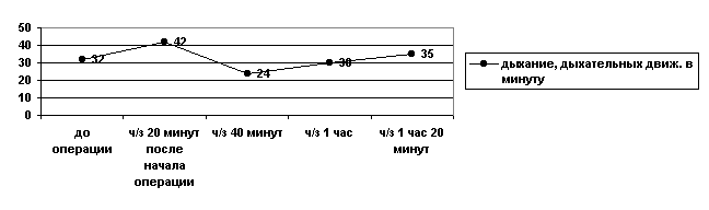
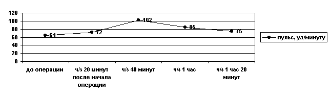
Dynamics of a pulse rate and respiration during operation
the Diary kuratsii
|
Date |
T, WITH |
P, ud /
Mines |
D,
Dyh/mines |
Flow of disease |
Therapy, a diet, a regimen of the content. |
|
3.03.2001 g |
38,6 |
75 |
32 |
The dog is depressed, respiration superficial, pulse rough. Morbidity in range of operation, a dog peeps. Began to drink water in the evening, the urination is kept. |
Rp.: Sol. Glucosae 5 %-100 ml
D. S. to a dog, intravenously, drop introduction.
*
Rp.: Sol. Acidi ascorbinici 5 % - 2 ml
D. t. d. N. 2 in amp.
S. to a dog, intravenously, drop introduction.
*
Rp.: Sol. Calcii chloridi 10 % - 5 ml
D.S. To a dog, intravenously, drop introduction.
*
Rp.: Sol. Sulfocamphocaini 10 % - 2 ml
D.S. To a dog, subcutaneously.
*
Rp.: Sol. Dimedroli 1 % - 1 ml
D. S. to a dog, unitary, intramuscularly.
*
Rp.: Sol. Analgini 25 % - 1 ml
D. S. to a dog, unitary, intramuscularly. |
|
4.03. |
37,8 |
67 |
29 |
Have removed the platen covering a suture. The dog rises, the defecation has appeared. Respiration equal, pulse of good filling. Appetite has appeared. Puffiness and a hyperemia in range of sutures. |
Rp.: Sol. Hydrogenii peroxidati 3 % - 50 ml
D. S. For processing a suture
*
Rp.: Sol. Viridis nitentis spirituosae 5 % - 10 ml
D. S. For processing a suture |
|
5.03. |
37,3 |
68 |
30 |
A condition of an animal without changes, the dog accepts I peep, is mobile. Presence of a small amount of a bloody exudate. The defecation and a urination are kept. |
To continue processing sutures.
Rp.: Sol. Viridis nitentis spirituosae 5 % - 10 ml
D. S. For processing a suture |
|
6.03. |
37,5 |
74 |
31 |
The dog is more awake, reduction of puffiness of range of operation is marked, morbidity at a palpation is insignificant. Ekssudatsija it is insignificant. |
Rp.: Sol. Viridis nitentis spirituosae 5 % - 10 ml
D. S. For processing a suture |
|
7.03. |
37,8 |
75 |
28 |
A condition of an animal satisfactory, in range of sutures crusts of a drying have appeared. The defecation and a urination are kept. |
Rp.: Lin. Synthomycini 10 % - 20,0
D. S. Outside, on range of sutures |
|
8.03. |
37,6 |
72 |
30 |
In range of operation the phenomena of a proliferation are marked. At a palpation puffiness, morbidity are absent. |
Rp.: Lin. Synthomycini 10 % - 20,0
D. S. Outside, on range of sutures |
|
9.03. |
37,8 |
68 |
29 |
Processes of a scarring in range of operation are marked. |
Rp.: Lin. Synthomycini 10 % - 20,0
D. S. Outside, on range of sutures |
|
10.03. |
37,5 |
68 |
28 |
The animal is mobile, awakely. Appetite good, accepts water in plenty. A defecation and a urination it is not broken. Pulse of average filling, equal, respiration rhythmical. On a place of an operational wound processes of a granulation are expressed. |
A removal of sutures. |
the Forecast:congenial for life of an animal.
the Outcome:convalescence.
the LIST of the USED LITERATURE- Anatomy of a dog. // Under B.M.Hromova's edition. Leningrad, "Science", 1972
- Veterinary preparations // the Manual under edition Tretjakova. M. IN "Agropromizdat", 1988 g, page 136.
- Danilevsky V.M.manual on veterinary therapy // М. "Kolos", 1983 year>
- General hirurgija. // Under V.Schmidt, V.Hartiga's edition, M.I. Cousines. 2 t. The joint edition of the USSR - GDR, "Medicine", 1985
- Operative surgery // Under I.I.Magdy's edition. Moscow, "Agropromizdat", 1990
- the Manual of a veterinary. Under edition Dostoevskogo of Items, Sudakova N.A. // Kiev, "Crop", 1990 g, page 437-438.
To discuss a material at a forum of a site
|